Publications
Recent advances in gene editing are enabling the engineering of cells with an unprecedented level of scale. But sorting large numbers of samples is laborious, and to date, no automated system exists to sequentially manage Fluorescence-Activated Cell Sorting (FACS) samples. Here, we describe the development of an integrated software and hardware platform to automate FACS, a central step for the selection of cells displaying desired molecular attributes. Our platform is built around a commercial instrument and integrates the handling and transfer of samples to and from the instrument, autonomous control of the instrument’s software, and the algorithmic generation of sorting gates, resulting in walkaway functionality. Automation eliminates operator errors, standardizes gating conditions by eliminating operator-to-operator variations, and reduces hands-on labor by 93%.
Liquid chromatography purification of multiple recombinant proteins, in parallel, could catalyze research and discovery if the processes are fast and approach the robustness of traditional, “one-protein-at-a-time” purification. Here, we report an automated, four channel chromatography platform that we have designed and validated for parallelized protein purification at milligram scales. The device can purify up to four proteins (each with its own single column), has inputs for up to eight buffers or solvents that can be directed to any of the four columns via a network of software-driven valves, and includes an automated fraction collector with ten positions for 1.5 or 5.0 mL collection tubes and four positions for 50 mL collection tubes for each column output.
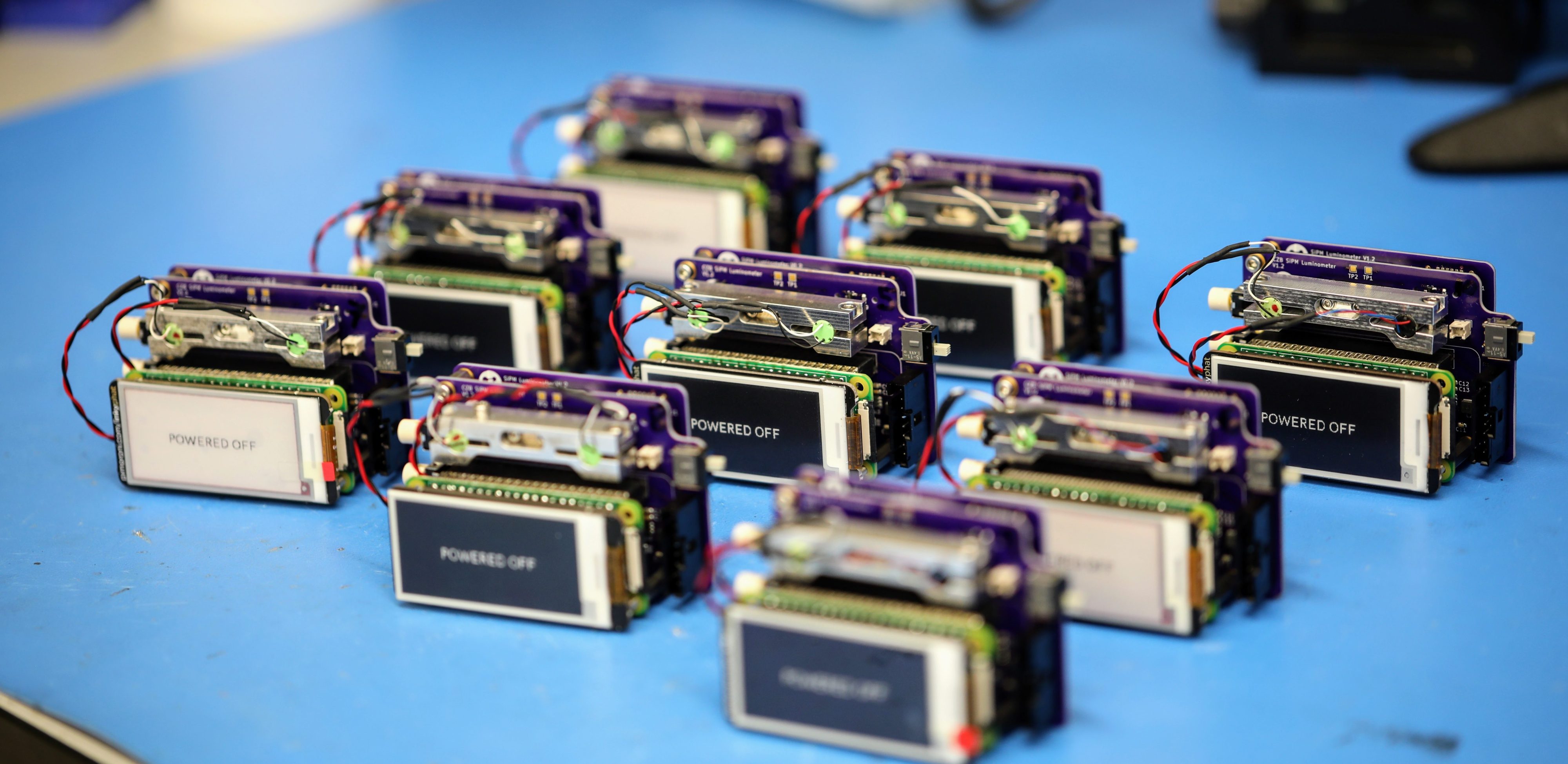
Luminescence is ubiquitous in biology research and medicine. Conceptually simple, the detection of luminescence nonetheless faces technical challenges because relevant signals can exhibit exceptionally low radiant power densities. Although low light detection is well-established in centralized laboratory settings, the cost, size, and environmental requirements of high-performance benchtop luminometers are not compatible with geographically-distributed global health studies or resource-constrained settings. Here we present the design and application of a ∼US $700 handheld, battery-powered luminometer with performance on par with high-end benchtop instruments. (Read a Twitter thread about the research here.)
Well-Lit: A programmable and customizable assistant for manual multi-well plate pipetting (Preprint)
We present here a device built to facilitate error-free pipetting of samples from individual barcoded tubes to a multi-well plate or between multi-well plates (both 96 and 384 wells are supported). The device is programmable, modular and easily customizable to accommodate plates with different form-factors, and different protocols. The main components are only a 12.3” touch screen, a small form-factor PC, and a barcode scanner, combined with custom-made parts can be easily fabricated with a laser cutter and a hobby-grade 3D printer. The total cost is between approximately US$550 and US$600, depending on the configuration.
Although qualitative rapid diagnostic tests (RDTs) and PCR-based assays for malaria detection have existed for many years, in most malaria-endemic countries manual counting by microscopy remains the dominant modality for assessment of infection. This method involves smearing, fixation, staining, and manual inspection of blood smears—a practice that is time-consuming, labor-intensive, error-prone, and has varied little in over a century. Similarly, for laboratories around the world that grow P. falciparum for research, the process of assessing different stages of parasite growth is a daily ritual. Here, we provide rigorous evidence that live, unstained parasites can be automatically distinguished and sub-categorized from a healthy background in the context of laboratory cell culture, by applying deep learning to ordinary microscopy images, with high-sensitivity and low false-positive rates.
Recent advancements in in situ methods, such as multiplexed in situ RNA hybridization and in situ RNA sequencing, have deepened our understanding of the way biological processes are spatially organized in tissues. Automated image processing and spot-calling algorithms for analyzing in situ transcriptomics images have many parameters which need to be tuned for optimal detection. Having ground truth datasets (images where there is very high confidence on the accuracy of the detected spots) is essential for evaluating these algorithms and tuning their parameters. We present a first-in-kind open-source toolkit and framework for in situ transcriptomics image analysis that incorporates crowdsourced annotations, alongside expert annotations, as a source of ground truth for the analysis of in situ transcriptomics images.
Measuring optical density (OD) is a common technique in biological laboratories to determine the concentration of a substance in solution or of bacteria (or microscopic particles) in suspension. For example, bacterial cultures engineered to produce (express) a protein or compound of interest are a workhorse of modern molecular biology laboratories. The most common tool for measuring OD is a spectrophotometer. However, most spectrophotometers are sophisticated, non-portable, and expensive laboratory instruments, costing tens of thousands of dollars. The problem is more acute in developing countries, where multiple labs have to share a single spectrophotometer, or there’s no such instrument at all. Here we present the detailed build instructions and characterization of a very simple OD meter that costs only US$60, and can measure OD values from ∼0.05 to 2.0.


