AI powers a better way to diagnose malaria
CZ Biohub San Francisco’s Remoscope shows promise in clinical study
After two years of working with his team to develop their malaria diagnostic device, Paul Lebel was nervous about testing it in a real-world setting — a clinic in Tororo, Uganda, an area with one of the highest malaria burdens in that country. The Remoscope can image and classify 2 million red blood cells as healthy or malaria-infected in just 12 minutes using a custom on-board neural network, and it had performed well at the San Francisco lab.
But how would it work in the field? What new challenges would they encounter?
“I told the team, I’m hoping we can achieve a limit of detection of under 500 parasites per microliter in the clinic,” he says, referring to a measure for the lowest concentration detectable in a patient’s blood. When infected red blood cells need to be correctly identified at levels less than 1 for every 10,000 healthy cells, it’s not an easy feat. Lebel wanted to keep his initial expectations in check.
That was January, 2023. Now, after a year of data collection and analysis, he was astounded by the results. “We were able to reliably detect 95 parasites per microliter in the clinical cohort using diluted blood, which is several times better than I expected,” says Lebel, the senior staff engineer who led the project within the Chan Zuckerberg Biohub San Francisco’s (CZ Biohub SF) Bioengineering team. “I was super happy with the result.”
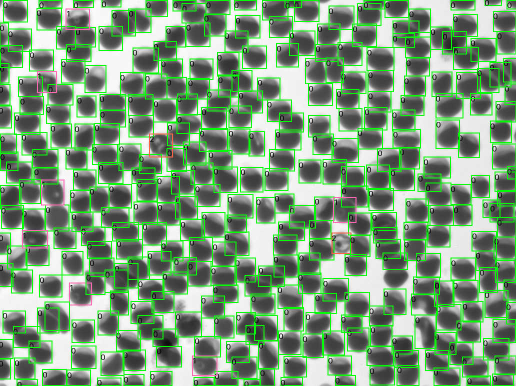
Conceived and developed at CZ Biohub SF, the Remoscope is a fully automated imaging cytometer that can image and count red blood cells, then use machine learning to classify cells as either healthy or harboring malaria parasites, all without the need for fixation or staining. Remoscope can even automatically classify infected cells by the parasites’ various life stages. Results from the field testing, in which Remoscope was used to analyze blood samples from more than 500 individuals in eastern Uganda, showed that it performed nearly as well as the conventional diagnostic technique, which has been in use for more than a century, and Lebel feels confident its accuracy can be improved.
A preprint of the clinical study is available on medRxiv. The study was facilitated by the Infectious Diseases Research Collaboration (IDRC), a nonprofit research organization in Uganda, and supported by the UCSF School of Medicine. Engineers and infectious disease specialists from the lab of Joe DeRisi, president of CZ Biohub SF, provided critical support and testing.
“What’s unusual about the San Francisco Biohub is that, from our founding, we’ve had engineers — people who know about electronics, optics, fluidics, manufacturing, and so on — working alongside our biologists,” says CZ Biohub SF Chief Technology Officer Rafael Gómez-Sjöberg, who also leads the Bioengineering team. “With this incredible combined expertise, over a two-and-a-half-year period or so, we developed a low-cost diagnostic solution — with imaging, AI, low-cost electronics, control software, and a custom disposable cartridge — then brought it to bear on the malaria parasite problem in the real world.”
The team hopes that commercial Remoscope devices will eventually be available for under $1,000.
“On our first try, we’re matching a technique that automation has been unable to improve upon in over 100 years,” says Lebel. “That’s not bad for your first shot, right?”
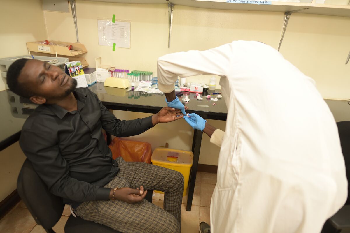
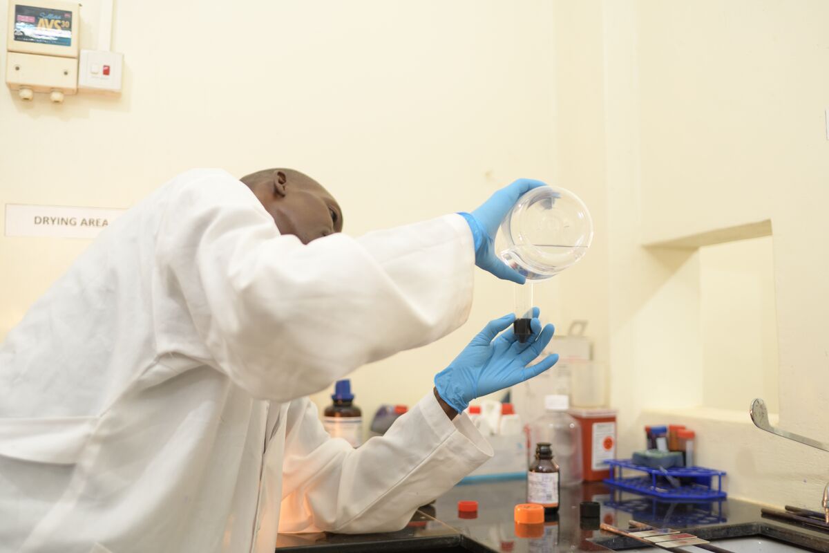
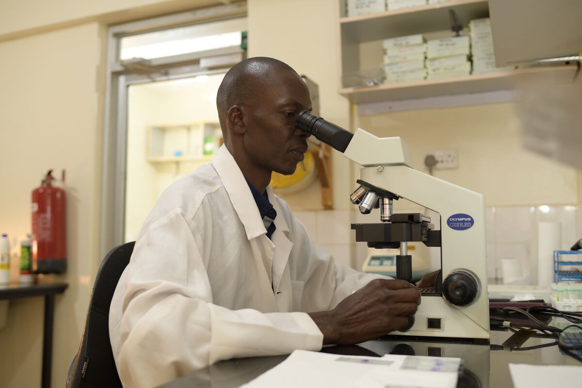
The technique of using a special stain to detect parasites in a blood smear was invented by the German chemist and bacteriologist Gustav Giemsa in the early 1900s, and microscopy with Giemsa staining has remained the gold standard for malaria diagnosis ever since. This approach requires a trained microscopist who visually scans a stained sample on a slide and manually counts infected cells. While it can be a reliable technique, results can vary widely depending on the skill, experience level, and fatigue of the microscopist, some of whom must analyze dozens of slides in a day. The quality of the reagents used can also be a factor in the accuracy of the results.
“One of the key learnings that the team took away from their trip to Uganda was how overburdened the personnel are in these centers that have high rates of malaria,” says DeRisi, who has studied malaria – and the parasite responsible for most cases of it – for more than 25 years. “There’s just not enough people during the day — not enough eyeballs or enough hours to diagnose all the slides and all the samples coming in. Many patients have to wait a day or more to know if they’ve been infected with malaria, and they may have had to return to their home by then, putting them out of reach for immediate treatment. A low-cost device like Remoscope that runs itself and runs at scale can vastly increase the throughput.”
A technician at the Infectious Diseases Research Clinic in Tororo, Uganda demonstrates the conventional method of malaria diagnosis by microscopy. (Credit: Celmo Media)
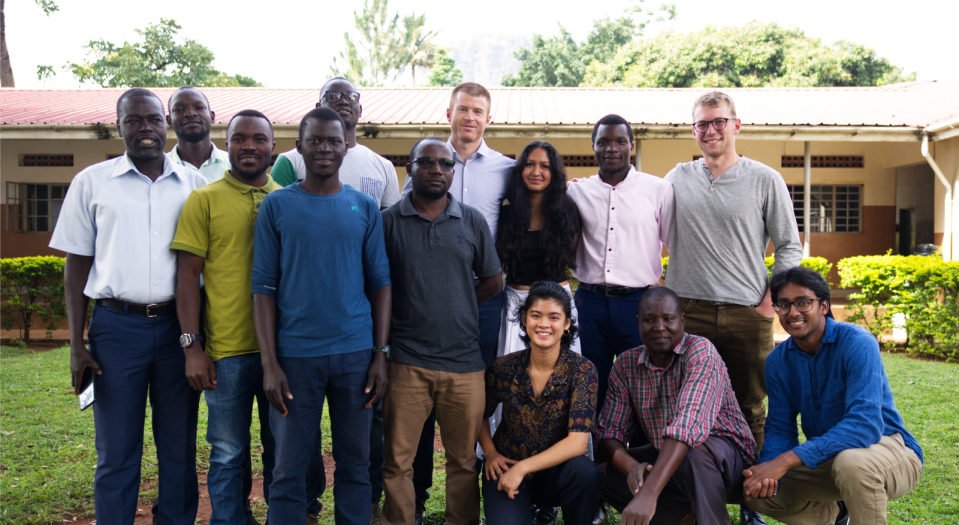
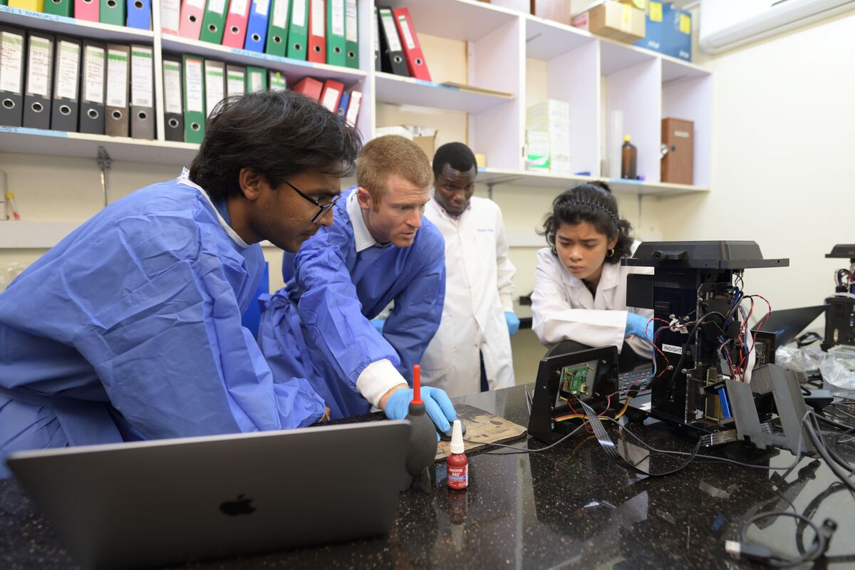
Three Remoscopes were shipped to the IDRC clinic in Tororo in late 2022 for launch of the clinical study. The San Francisco team visited Uganda last year, spending a week with IDRC employees James Emorut and Peter Olwoch to check in on the study and to upgrade the devices.
Biohub engineers (from left) Ilakkiyan Jeyakumar, Paul Lebel, and Michelle Khoo (far right), with IDRC technician James Emorut (second from right) work on Remoscope circuitry at the IDRC clinic in Tororo, Uganda. (Credit: Celmo Media)
“They ran a high-quality clinical study,” Lebel says. “Their expertise was essential.”
In trying to answer the question of how the Remoscope’s diagnostic accuracy compares to that of Giemsa (thick blood smears), the answer is that it depends on the microscopist. Specifically, the Remoscope’s limit of detection was found to be 95 parasites per microliter. “A really good technician can sometimes succeed at finding 15 parasites per microliter, but that’s uncommonly good,” says Lebel. “It’s like saying humans can run 100 meters in under 10 seconds, when there’s really only an elite few people in the world who can do that. In practice, less than one-third of microscopists globally detect parasites correctly at even 100 parasites per microliter.”
In that sense, Remoscope surpassed the global average for Giemsa in the limit of detection. But compared to studies in which pairs of highly trained expert technicians double-checked samples, Remoscope trailed slightly.
The other two important metrics of accuracy are sensitivity, or how often a test returns a false negative, and specificity, or how often there is a false positive. “The Remoscope is equally sensitive as expert-level microscopy, but not quite as specific yet, but we think it will be,” Lebel says.
The reason for his confidence is that the team has already seen improvements in specificity by moving from diluted blood samples to undiluted blood. “With whole, undiluted blood it’s a cleaner result and there are fewer false positives,” Lebel says, adding that these results were achieved in the lab, but that data on undiluted blood from the field testing in Uganda is still being collected.
The Biohub team named the device after the Acholi word for blood, which is “remo” (pronounced like “demo”); the Acholi are an ethnolinguistic group who live in northern Uganda.
The Remoscope is a diminutive device, just under 1 foot tall (300 mm, to be precise), with a 7-inch touchscreen. The cost to build a lab prototype is about $2,000, and the cost to run a test is about $1, which is the cost of the disposable flow cell that holds the blood sample. “We’re the only automated test that matches the price of Giemsa,” Lebel says.
The software is trained to recognize four life-cycle stages of Plasmodium falciparum, the parasite responsible for 90% of malaria deaths worldwide, in red blood cells, including ring, trophozoite, schizont, and gametocyte. Lebel says it should be possible to train the algorithm to recognize other malaria species as well, but that has not yet been tested.
The device is able to image and classify 2 million cells in 12 minutes, meaning the test result can be available in as little as 1 minute, compared to at least 45 minutes for Giemsa staining.
“We’ve been doing a lot of research in HIV, TB, and malaria,” says Olwoch, the IDRC lab manager for the past 12 years. “Over the years, we have tested a few automated microscopes with different functionalities. Most of them would use stained blood preparations that require the use of reagents that stain for a long period. And the turnaround time is a minimum of one hour. That’s time we can use for clinical management and treatment of the patient. It’s way too much.”
Olwoch was pleased not only by Remoscope’s speed but also that it could allow the clinic to eliminate the need for reagents, which can be expensive, and certain pieces of equipment.
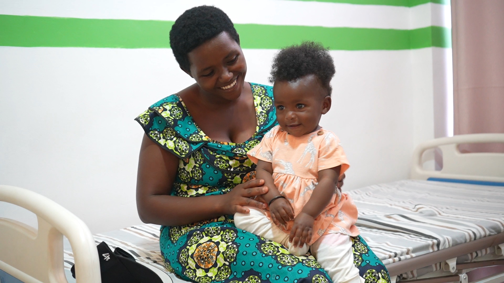
The Remoscope team is continuing to work with IDRC to evaluate testing with undiluted blood and other improvements that will lead them to scaling up. Separately, the team has struck up a collaboration with Goodlife Access, an independent California-based initiative working on healthcare issues in Rwanda, to study how the device performs in Rwanda. They will examine not only the diagnostic performance but also practical and economical outcomes.
Beyond malaria, the Remoscope engineers are exploring additional applications for the device, which in the future could include detecting other bloodborne pathogens, genetic blood disorders, blood cancers such as leukemia, and more.
Meanwhile, the team is actively seeking partners who can help scale up the manufacture and distribution of the device, getting it to the locations where it’s most needed. “The most important thing for treating malaria is knowing who has the parasite, and who doesn’t, so accurate and fast diagnosis will help to quickly triage patients into appropriate treatment, instead of waiting potentially a day or more, when mosquitos can feed on that person and continue the cycle of infections,” says DeRisi. “Rapid treatment is required to break the chain.”
A conversation with Steve Quake, Amy Herr, and the Stellar Science Foundation’s Takanori Takebe
Learn More
A ‘Google Earth’ of embryology, CZ Biohub San Francisco's zebrafish cell atlas brings a new vision to developmental biology
Learn More
CZ Biohub Chicago’s implantable device demonstrates an impactful new way to monitor health and disease
Learn More
Stay up-to-date on the latest news, publications, competitions, and stories from CZ Biohub.
Marketing cookies are required to access this form.