Everything everywhere all at once: 'Zebrahub' tracks development like never before
A ‘Google Earth’ of embryology, CZ Biohub San Francisco's zebrafish cell atlas brings a new vision to developmental biology
When early cartographers undertook perilous expeditions to map unknown corners of the world with sextants, compasses, and hand-drawn diagrams, it’s unlikely they imagined that someday anyone with an internet connection would have access to a seamless view of the entire planet from the comfort of their own home. Today, pioneering scientists are working to create a similar experience for a much tinier, but no less important domain: developing embryos. The goal is to track and map the behavior of each and every cell working together to create an adult lifeform, and present that map in a clickable, navigable display — a sort of Google Earth for developmental biology.
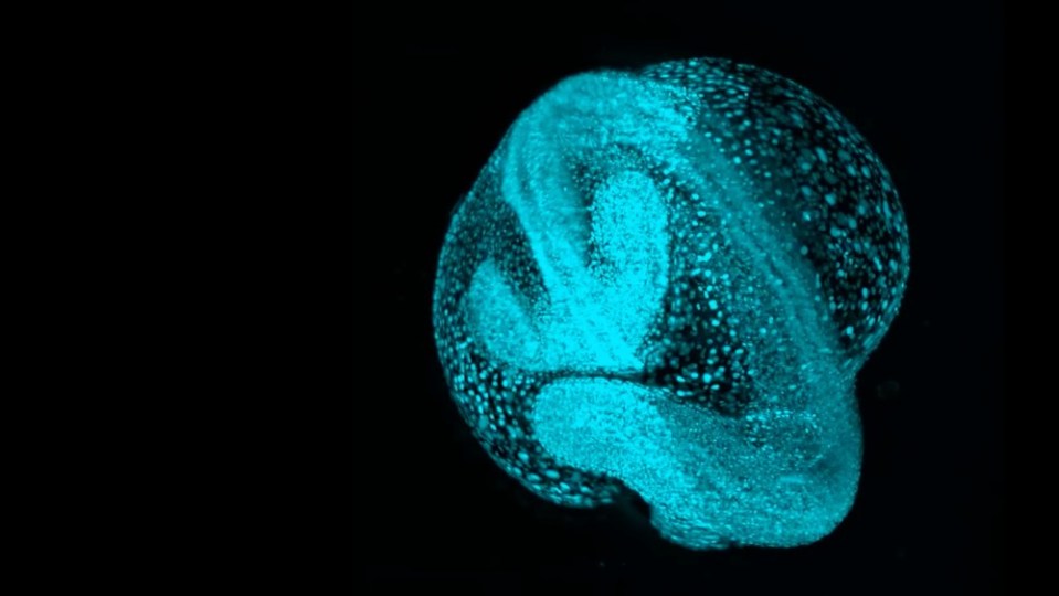
Now, in a new paper in Cell, researchers at the Chan Zuckerberg Biohub San Francisco (CZ Biohub SF) are unveiling the latest advancements in that effort. “Zebrahub” is a state-of-the-art cell atlas that combines high-resolution time-lapse videos of newly emerging cells in zebrafish embryos with extensive data on which genes are switched on and off as individual cells navigate to their eventual stations and “decide” what role they will ultimately play in the body of the adult fish.
A freshwater species native to South Asia, zebrafish as adults rarely exceed two inches in length, and they are a long-established model for developmental research relevant to human health. Around 70% of human genes have counterparts in zebrafish and, though we look quite different, as fellow vertebrates we share most of the same overall body plan in addition to the cellular and molecular processes by which various body parts initially form. Critically, zebrafish embryos are also mostly transparent and — unlike those of, say, mice — develop outside the mother, making it possible for scientists to observe their early growth in detail under a microscope.
Creating Zebrahub, which is free to all and includes built-in analytical tools designed for biologists, required building a suite of new instruments and software. It’s the most comprehensive atlas of its kind and, as the researchers write in the paper, an important step towards “ushering in a new era for developmental and evolutionary biology.”
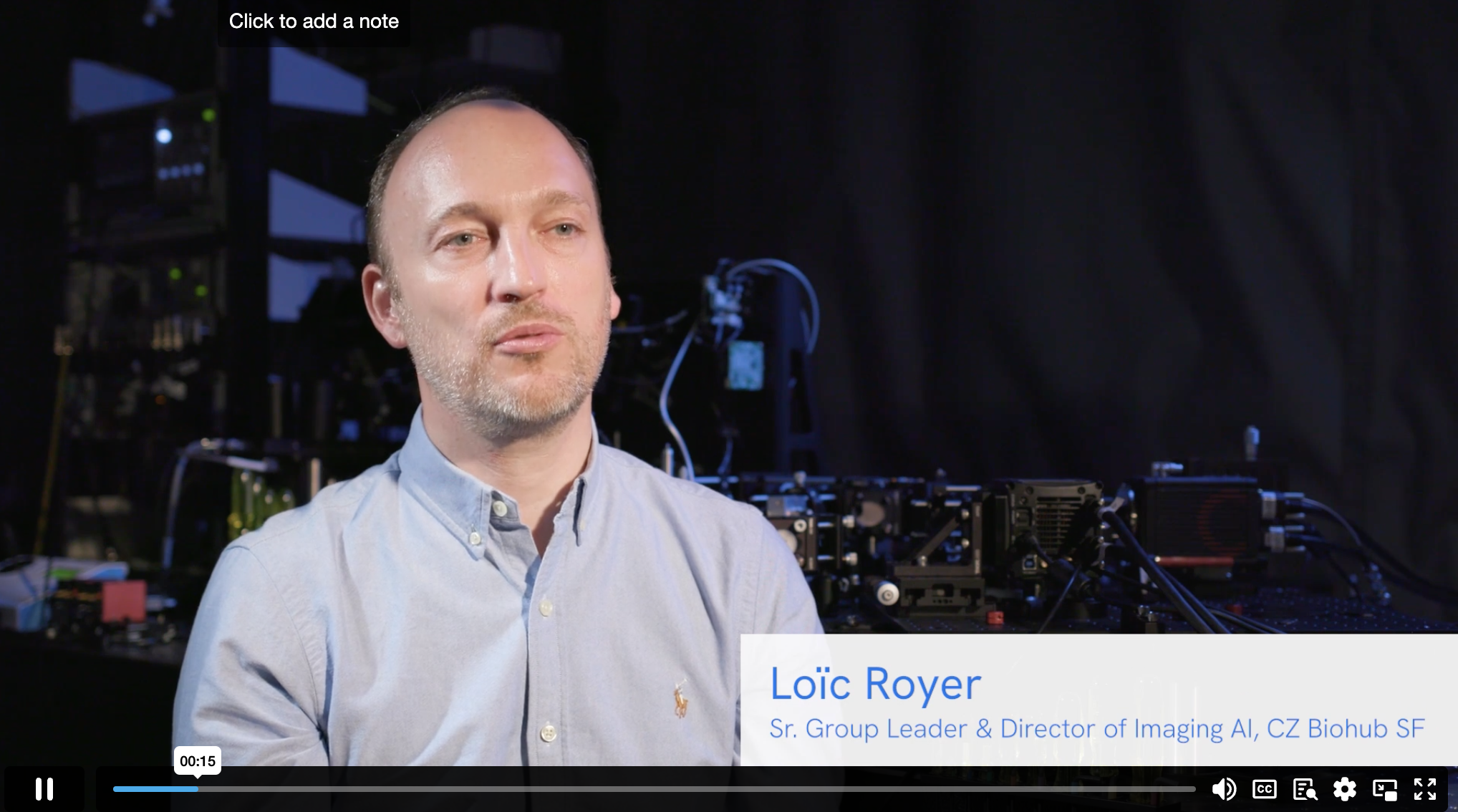
“How a lifeform goes from a single cell to an entire body is one of biology’s biggest mysteries,” says senior author Loïc Royer, leader of the Organismal Architecture group and director of imaging AI at CZ Biohub SF. “With Zebrahub, we’ve created possibly the most detailed map of that process ever.”
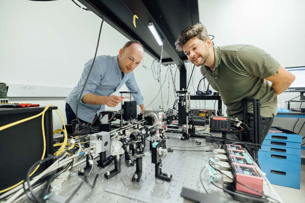
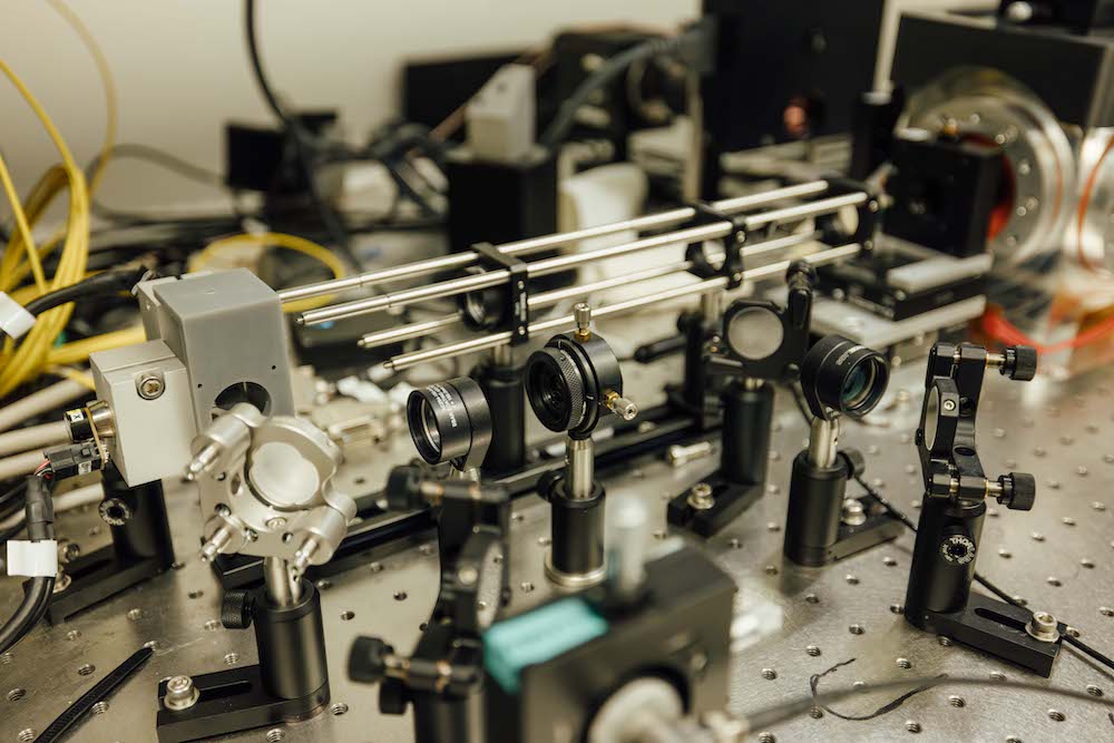
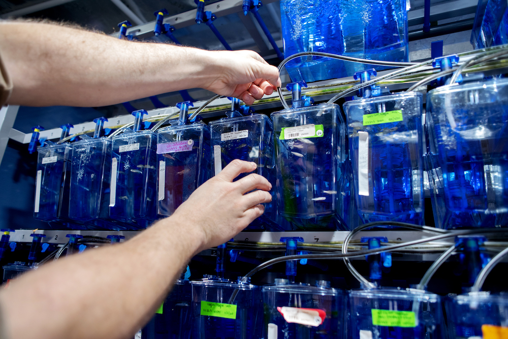
CZ Biohub SF scientists Loïc Royer (left) and Merlin Lange work on a light-sheet microscope (top right) designed and built at the Biohub. They use it to study zebrafish embryos; the fish are kept in the “zebrafish hotel” (bottom right) at Biohub’s labs. | Photos by Dale Ramos (left, top right) and Barbara Ries (bottom right).
To form a complex adult organism such as a human or fish, a fertilized egg must split into a set of progeny that continue dividing until millions of cells have been born and have assumed their roles as parts of the skin, liver, brain, and all the other components of the body. While, for the most part, all cells of an embryo contain an identical set of genes, the way that each type of cell uses these genes — switching them on and off in different combinations at different timepoints — is unique. Scientists have long pondered just how the “choices” regarding thousands of genes in millions of cells come together to create a fully functioning adult lifeform with many types of specialized tissues. Each advancement towards solving this daunting puzzle yields new insights about why the process sometimes goes wrong, leading to disorders and disease.
But even with powerful models like zebrafish, developmental biology has historically been conducted in a piecemeal fashion, limited by the complexity of examining events far too tiny to see, and happening by the millions across the bodies of fragile living organisms that can be easily damaged by the very experiments designed to understand them. As in the early days of cartography — before satellites snapped pictures of Earth from space and cars with spinning cameras mapped our streets — the field has made its breakthroughs in fits and starts, and has lacked a comprehensive system for considering the whole instead of just the pieces. With Zebrahub, researchers at CZ Biohub SF hope to help change that, accelerating the field by giving researchers easy access to the breadth of these processes, all in one place.
Thanks to a new set of laboratory procedures developed at CZ Biohub SF, Zebrahub is also one of the first datasets of its kind to include gene expression data specific to individual embryos, as the process of collecting such data has typically required researchers to pool DNA from multiple embryos together. This means Zebrahub confers the added benefit of allowing scientists to investigate the subtle expression differences that might give rise to different health outcomes among sibling fish.
“Zebrahub offers one of the first opportunities to investigate the behavior of cells in the extremely complex process of development with extremely high precision,” says Merlin Lange, a CZ Biohub SF senior staff scientist and first author of the new Cell paper. “It’s very rare to combine both gene expression from individual cells and spatial mapping of cells over time in the same resource like this.”
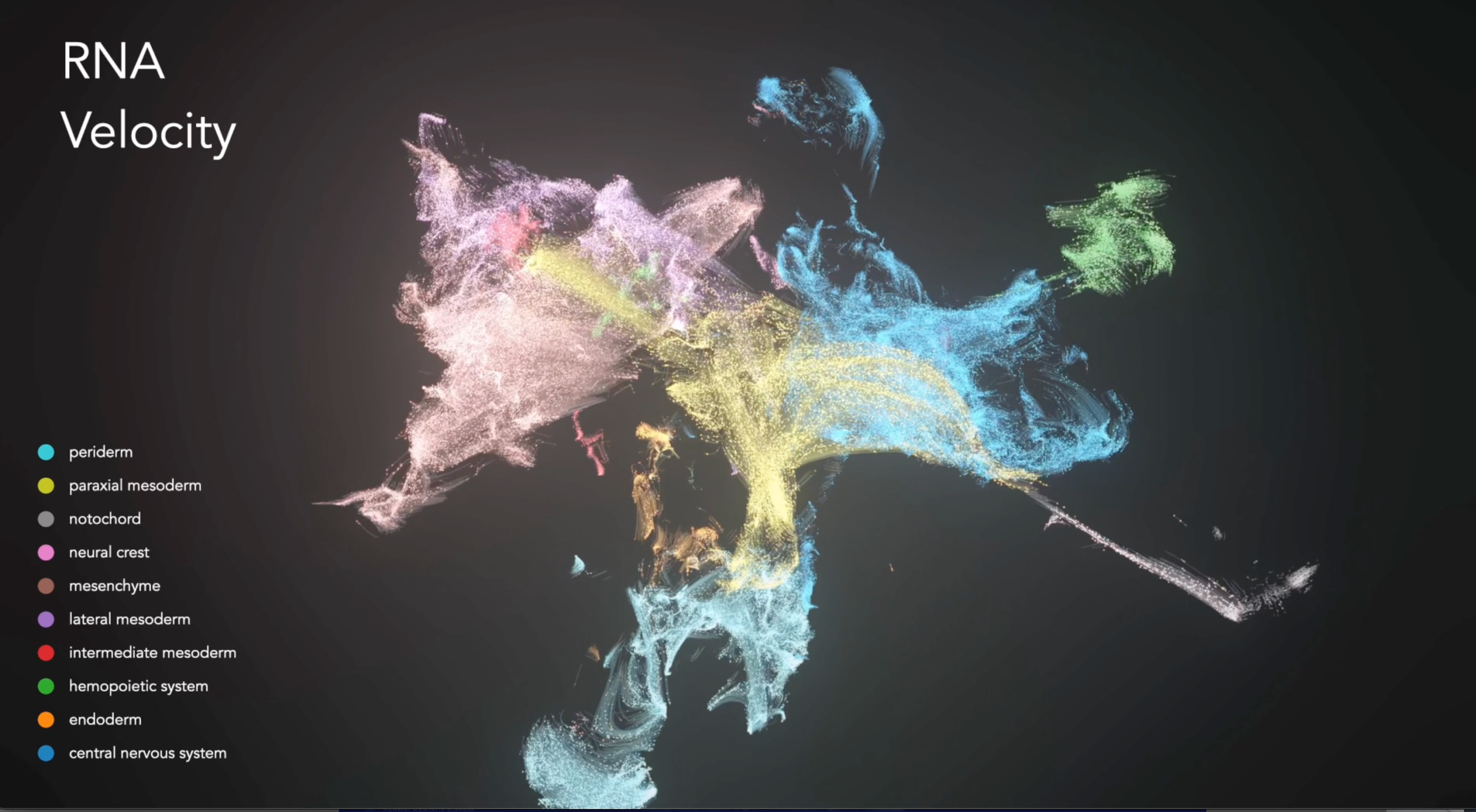
Zebrahub features two major datasets, along with a suite of tools designed to help biologists use them. The first offers time-lapse video microscopy showing the birth and early movements of most cells in a zebrafish embryo in the first 24 hours after fertilization, during which time organs start to form. The second provides data on which genes were active in more than 120,000 zebrafish cells at 10 separate time points during the embryos’ first 10 days.
To create the time-lapse videos, Royer, Lange, and CZ Biohub SF scientists and engineers designed and built “DaXi” (pronounced “dah-shee”), a new kind of automated microscope with a field of view large enough to capture images of entire living embryos. DaXi is a so-called light-sheet microscope that emits and captures light in a unique way designed to protect embryos from high-intensity laser beams that would damage or even kill the embryo after a short period of time.
Then, to allow scientists to easily use the captured videos to study specific cells, CZ Biohub SF software engineer Jordão Bragantini led the development of a sophisticated new program called Ultrack, which automatically identifies cell nuclei (typically the most distinctive landmark in a cell) and tracks their movements in the videos over time in three-dimensional space. Combined, the datasets generated by these tools allow researchers to conduct “virtual experiments” examining where cells begin and end up during development — even running their developmental trajectory backward and forward in time.
In just developing this methodology, the Zebrahub team has already made some intriguing discoveries. For example, the team looked at a subset of cells in the embryo’s tail called neuro-mesodermal progenitors, which, at the timepoints they examined, had previously been thought to only be able to give rise to one type of tissue. However, as the Zebrahub researchers analyzed the cells’ movement and expansion, they realized these cells were actually developing into both muscle cells and neurons that were integrating into the spinal cord.
“This was a very unexpected finding,” Lange says. “And it’s the kind of thing that would be hard to confirm without the broad view that Zebrahub provides.”
Zebrahub, which has been available online to researchers for just over a year, has already helped support discoveries from other labs. One team that included researchers from Ashland University in Ohio and the State University of New York in Albany used Zebrahub in concert with their own cell atlas to ask which cellular proteins might contribute to the formation of cataracts in the eye. For this, the researchers relied on Zebrahub’s gene expression database to see when the cells of the lens activate and deactivate certain genes in a way that might lead to problems.
“Zebrafish are really small, and it’s really difficult for us to peel the lens apart in order to ask questions about what genes are working in this region and how one cell might be different from another,” says Mason Posner, a professor of biology at Ashland and co-senior author of the study. Here, “that’s already been done for us and we can get these deep understandings about, for example, how this tissue even becomes transparent and functions, essentially, as biological glass.”
A project five years in the making, Zebrahub required the development of numerous new technologies to achieve and relied on experts in the fields of biology, engineering, optics, physics, and data science housed under the roof of CZ Biohub SF. Every piece of technology developed in the process is open-source, which will contribute to more data being added to the project as the community works together to improve our view of embryo development.
“This kind of project would never be funded through conventional channels,” says Sandra Schmid, chief scientific officer of CZ Biohub SF. “But thanks to the Chan Zuckerberg Initiative’s innovative approach to building scientific institutes, Zebrahub has not only provided millions of measurements about embryonic development that anyone in the scientific community can access, it’s also exactly the kind of data needed to power new AI initiatives that stand to take us into the future of health science.”
CZ Biohub Chicago’s implantable device demonstrates an impactful new way to monitor health and disease
Learn More
For CZ Biohub San Francisco research associate Rodrigo Baltazar-Nuñez, science is personal
Learn More
In the last couple decades, Malaysia has emerged as a global hotspot for neuroinfections such as encephalitis. Now a team from Universiti Malaya is...
Learn More
Stay up-to-date on the latest news, publications, competitions, and stories from CZ Biohub.
Marketing cookies are required to access this form.